Introduction
Supernumerary teeth are a relatively frequent disorder of odontogenesis characterized by the presence of tooth in addition to the normal series. Supernumerary or extra teeth constitute approximately 15% of teething anomalies. They are often found in the area of incisor teeth in the maxilla, second premolar teeth in the mandible and maxilla, and molar teeth.Extra teeth may present in both permanent and primary dentitions, but are five times less frequent in the primary dentition. The literature reports that 80 to 90% of all supernumerary teeth occur in the maxilla, of which half are found in the anterior region . The lateral incisor is the most frequently observed supernumerary tooth in primary dentition.[1]
The most common type of supernumerary tooth as indicated by Alberti[2] is mesiodens accounting for 80% of all supernumerary teeth[1]. It is found mainly in the premaxilla between the two central incisorsand rarely in the mandible. Mesiodens may occur as single, mutiple, unilateral or bilateral. They can, On the basis of its morphology, be supplemental, conical, or tuberculate[3],[4]. The presence of multiple supernumerary teeth is called 'mesiodentes'[5].
various studies have reported the incidence of mesiodens between 0-1.9% for deciduous teeth and between 0.15-3.8% for permanent teeth with male to female occurrence ratio of 2:1[6],[7].Reports of supernumerary teeth are quite common in dental literature. These are discovered on a complaint by a patient or when the patient seeks treatment for malocclusion.
Congenital lack of one or more tooth is another common anomaly, mostly involving second premolars and maxillary lateral incisors. Terms like hypodontia, oligodontia and anodontia have been used to describe various presentations of this anomaly. It can be unilateral or bilateral, but reports of bilateral missing teeth are rare.
The present case reportdescribes an unusual association of two dental anomalies: a rare presentation of mandibular mesiodens and bilateral congenital absence of the mandibular central incisors.
Case Report
A 22 year-old male reported with a complaint of dirty teeth and bleeding gums. Intraoral examination revealed a mesiodens between the permanent mandibular lateral incisors with congenitally missing cental incisors (Fig. 1). The coronal portion of the mesiodens exhibited conical shape. Spacing was observed between lateral incisors and mesiodens.
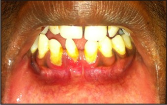 | Fig 1 : Mesiodens Between The Permanent Mandibular Lateral Incisors With Congenitally Missing Cental Incisors
 |
Intraoral periapical radiograph revealed a single conical root with a single root canal with absence of cental incisors (Fig. 2).
 | Fig 2 : Intraoral Periapical Radiograph Revealed A Single Conical Root With A Single Root Canal With Absence Of Cental Incisors
|
No other teeth were missing. Soft tissues were normal. There was no relevant medical and family history and the patient was otherwise healthy.
The patient was normal in his facial appearance, did not exhibit any physical or skeletal abnormality and showed no signs of mental retardation. No such anomaly was observed in his two sisters and parents when called for examination.
As the patient was asymptomatic and was not bothered about spacing, no treatment other than oral prophylaxis was rendered.
Discussion
Supernumerary teeth are a developmental disturbance occurring during odontogenesis resulting in the formation of teeth in excess of the normal number. They occur both in the deciduous and permanent dentition.
The first report of a supernumerary tooth appeared between AD 23 and 79[8].The first documented report of supernumerary teeth has been found in the ancient human skeletal remains of the Lower Pleistocene era. Until recently, the most primitive evidence of the presence of mesiodens goes back to 13,000 years, when it was found among the remains of an Australian aborigine[9]. Balk (1917) defined mesiodens as the most common among supernumerary teeth, located mesial to both central incisors; appearing peg shaped, in a normal or inverted position[10]. Regezi and Sciubba[11] mentioned that the anterior midline of the maxilla is the most common site of the supernumerary tooth, hence the supernumerary tooth is known as mesiodens. Very few supernumerary teeth have been reported in the primary dentition[12].
The etiology of the supernumerary teeth is not completely understood. It was originally postulated that mesiodens represented a phylogenetic relic of extinct ancestors who had three central incisors[13]. The presence of supernumeraries in family members suggests heredity as an etiological factor; however, it does not follow a simple Mendelian pattern. Autosomal dominant inheritance with incomplete penetration has been the proposed genetic theory. A sex-linked pattern has also been proposed as males are affected twice as frequently as females[14]. A second theory known as dichotomy suggests that the tooth bud is split to create two teeth, one of which is the mesiodens[15]. The third theory involving hyperactivity of the dental lamina is the most widely supported. According to this theory, remnants of the dental lamina or palatal offshoots of the active dental lamina are induced to develop into an extra tooth bud, which results in a supernumerary tooth[16]. Supernumerary teeth may occur as a single isolated anomaly or in association with syndromes like cleft lip and palate, Downs syndromes, Cleidocranial dysplasia, etc[17].
Few studies on the prevalence of mesiodens involving certain ethnic or racial populations have been published to date. The prevalence of mesiodens has been estimated to be 0.45% in Caucasians, 0.4% in Finnish, 1.43%in Norwegians, 2.2% in Hispanic populations, and 8.3% in a group of Turkish children. In a study by Kaan, the prevalence in Turkey has been found to be 0.3%[18]. The prevalence of supernumerary teeth in the permanent dentition of the Caucasian general population has been reported to be between 0.1 and 3.8%. The estimated prevalence in the sub-Saharan Africa and Asian population is reported to be between 2.7 and 3.4%. The general prevalence of mesiodens in Iranian children has been seen to be 1.6%, as reported by Mieghani[19].
Supernumerary teeth are usually classified on the basis of their occurrence in the permanent dentition (rudimentary mesiodentes) or the primary dentition (supplementary mesiodentes) andtheir morphology together with their location in the dental arches.According to the shape and size, two subclasses are considered in the classification of mesiodens; namely, eumorphic and dysmorphic. The eumorphic subclass is usually similar to a normal-sized central incisor, whereas, the dysmorphic teeth have different shapes and sizes and are categorized into conical, tuberculate and molariform[20]. Conical mesiodens are the most commonly observed and appear as peg-shaped and are located palatally between the maxillary central incisors. They have a completely formed root and can erupt into the oral cavity. However, they may also be inverted with the crown pointing superiorly in which case they are less likely to erupt into the oral cavity.Tuberculate mesiodens are barrel-shaped with several cusps or tubercles and have incomplete or abnormal root formation. They rarely erupt into the oral cavity. A much rarer type of mesiodens is the molariform mesiodens, which has a premolar-like crown and a completely formed root[21].
Microscopically, changes have been observed in the structure of dentine and enamel of supernumerary teeth - mesiodenses. The highest incidence of changes was observed in radicular dentine, where tubules in a vestigial form were found suggesting inhibited development of mesiodens radicular part[22].
A mesiodens may erupt normally, stay impacted, appear inverted or take a horizontal position. Only 25% of all mesiodentes spontaneously erupt into the oral cavity. Asymptomatic unerupted mesiodens may be discovered during radiological examination of the premaxillary area[23]. Kaan found 37.6% in the inverted position and 7% in a horizontal position in a radiographic study[18]. The presence of mesiodens often results in complications including, retention of primary teeth, delayed eruption and ectopic eruption of permanent teeth, closure of the eruption path, rotations, retention, root resorption, pulp necrosis, crowding, and diastema, as well as nasal eruption and formation of dentigerous and primordial cysts[1],[4]. According to Tashima, the prevalence of inter-incisal diastema is seven times higher in the presence of mesiodens[24]. Therefore, early detection and management of all supernumerary teeth becomes a necessary part of preventive dentistry. However, symptomless cases could be left untreated along with regular check-ups. Less common complications involving the permanent incisors include dilacerations of the developing roots and loss of tooth vitality. Therefore, early diagnosis of mesiodens has particular importance in terms of preventing such complications.
There is limited evidence indicating mesiodens as a risk factor in trauma. The only case suggesting mesiodens as a risk factor was reported by Kupietzky[25] and more recently by A. Alacam, where the mesiodens was considered as a risk factor for causing as well as complicating dental trauma[26].
Hypodontia is the congenital absence of less than six teeth because of agenesis. The most frequently occurring congenitally missing permanent teeth, apart from third molars, are the mandibular second premolar (3.4%) and the maxillary lateral incisor (2.2%)[27]. The absence of teeth may be unilateral or bilateral. There are reports showing unilateral occurrence of permanent mandibular central incisors[28]. But agenesis of bilateral mandibular central incisors is not well documented. The first report of congenitally missing two mandibular incisors was given by Newman in 1967[29]. It has been reported that missing mandibular incisors is common in certain populations like Japanese, Korean and Chinese[30].
Although the exact etiology of congenital agenesis of both central incisors is unknown, but it has been attributed to several factors like trauma, radiation, infection, metabolic disorders and idiopathic changes[31]. Newman and Newman have given four main theories mainly for the cause of agenesis of incisors. Heredity or familial distribution is the primary cause. Second, anomalies in the development of the mandibular symphysis may affect the dental tissues forming the tooth buds of the lower incisors. Third, a reduction in the dentition regarded as nature’s attempt to fit the shortened dental arches (an expression of the evolutionary trend) and finally, localized inflammation or infections in the jaw and disturbance of the endocrine system destroying
the tooth buds[28]. It has also been reported that genes MSX1, TGFA and PAX9 interaction sometimes play a role in human tooth agenesis[32].
The present case is rare due to a rare combination of two dental anomalies was observed in mandible with presentation at a much rarer location. The medical and family history ruled out any association with any syndrome or genetic abnormality.Only one such case has been published in literature till date.
Conclusion
Hypodontoia and supernumerary teeth in permanent dentition are common anomaliesaffecting the normal development of dentition. Early diagnosis of these anomalies is important for the preservation of the dentition and the development of the occlusion.
References
1. Ferrĕs-Padr!5;E, Prats-Armengol J, Ferrĕs -Amat E. A descriptive study of 113 unerupted supernumerary teeth in 79 pediatric patients in Barcelona. Med Oral Patol oral cir Bucal 2009;14:E146-52.
2. Alberti G, Mondani PM, Parodi V. Eruption of supernumerary permanent teeth in a sample of urban primary school population in Genoa, Italy. Eur J Paediatr Dent 2006;7:89-92.
3. Garvey MT, Barry HJ, Blake M. Supernumerary teeth -an overview of classification, diagnosis and management. J can Dent Assoc 1999;65:612-6.
4. Primosch RE. Anterior supernumerary teeth -assessment and surgical intervation in children. Pediatr Dent 1981;3:204-15.
5. Gallas MM, García A. Retention of permanent incisors by mesiodens: A family affair. Br Dent J 2000;188:63-4.
6. Prabhu NT, Rebecca J, Munshi AK. Mesiodens in the primary dentition: A case report. J Indian Soc Pedo Prev Dent 1998;16:93-5.
7. Van Buggenhout G and Bailleul-Forestier I., Eur J Med Genet 51, 178-81 (2008)
8. Maya C, Ashok Kumar BR. Familial occurrence of mesiodens with unusual findings: Case report. Quintessence Int 1998;29:49-51.
9. Sutton PR. Tooth eruption and migration theories: Can they account for the presence of a 13,000-year-old mesiodens in the vault of the palate? Oral Surg Oral Med Oral Pathol 1985;59:252-5.
10. Jiau Fu Z, Mauricio M, David LK, Robert JH. Supernumerary and congenitally absent teeth: A literature review. J Clin Pediatr Dent 1996;20:87-95.
11. JA Ragezi, JJ Sciubba. Oral Pathology - Clinical Pathologic Correlation. 3rd ed. Philadelphia: London, New York: W. B. Saunder Co.; 1999.
12. Luten JR Jr. The Prevalence of supernumerary teeth in primary and mixed dentition. J Dent Child 1967;34:346-53.
13. Von Arx T. Anterior maxillary supernumerary teeth: A clinical and radiographic study. Aus Dent J 1992;37:189-95.
14. Ersin NK, Candan U, Alpoz AR, Akay C. Mesiodens in primary, mixed and permanent dentitions: A clinical and radiographic study. J Clin Pediatr Dent 2004;28:295-8.
15. Sedano HO, Gorlin RJ. Familial occurrence of mesiodens. Oral Surg Oral Med Oral Pathol 1969;27:360-1.
16. Primosch RE. Anterior supernumerary teeth-assessment and surgical intervention in children. Pediatr Dent 1981;3:204-15.
17. Zhu JF, Marcushamer M, King DL, Henry RJ. Supernumerary and congenitally absent teeth: A literature review . J clin pediatr Dent 1996;20:87-95.
18. Gündüz K, Celenk P, Zengin Z, Sümer P. Mesiodens: A radiographic study in children. J Oral Sci 2008;50:287-91.
19. Meighani G. Prevalence of Mesiodens in Iranian children. A Radiographic Study. Iran J Orthod 2009;1:31-6.
20. Van Buggenhout G, Bailleul-Forestier I. Mesiodens. Eur J Med Genet 2008;51:178-81.
21. Sharma A, Gupta S, Madan M. Uncommon mesiodens: A report of two cases. J Indian Soc Pedo Prev Dent 1999;17:69-71.
22. Kim SG, Lee SH. Mesiodens: A clinical and radiographic study .ASDC J Dent child 2003;70:58-60.
23. Liu JF. Characteristics of premaxillary supernumerary teeth: A survey of 112 cases. ASDC J dent child 1995;62:262-5.
24. Tashima AY, Alencar CJF, Fonoff RN, Wanderley MT, Haddad AE. Correlation between the prevalence of supernumerary teeth and its consequences for the development of the occlusion. J Appl Oral Sci 2007;15:34.
25. Kupietzky A, Rotstein I, Kischinovsky D. A multidisciplinary approach to the treatment of an intruded maxillary permanent incisor complicated by the presence of two mesiodentes. Pediatr Dent 2000;22:499-503.
26. Alaçam A, Bani M. Mesiodens as a risk factor in treatment of trauma cases. Dent Traumatol 2009;25:25-31.
27. Bäckman B and Wahlin YB Variations in number and morphology of permanent teeth in 7- year-old Swedish children. Int J Paediatr Dent 2001;11(1): 11-17.
28. Newman GV and Newman RA. Report of four familial cases with congenitally missing mandibular incisors. Am J Orthod Dentofacial Orthop 1998; 114(2): 195-207.
29. Newman GV. Congenitally missing mandibular incisors: treatment procedures. Am J Orthod 1967; 53(7): 489-491.
30. Davis PJ. Hypodontia and hyperdontia of permanent teeth in Hong Kong schoolchildren. Community Dent Oral Epidemiol1987; 15(4): 218-220.
31. Endo T, Ozoe R, Kubota M, Akiyama M and Shimooka S. A survey of hypodontia in Japanese orthodontic patients. Am J Orthod Dentofacial Orthop 2006; 129(1): 29-35.
32. Vieira AR, Meira R, Modesto A and Murray JC. MSX1, PAX9, and TGFA contribute to tooth agenesis in humans. J Dent Res 2004; 83(9): 723-727.
|